As development progresses, small lacunae begin to form within the extraembryonic mesoderm which enlarge to become the extraembryonic coelom. 1994;83(5 Pt 1):647-651. Thegestational sac (GS) is the first sign of early pregnancy on ultrasound and can be seen with endovaginal ultrasound at approximately 3-5 weeks gestation when the mean sac diameter (MSD)would approximately measure 2-3 mm in diameter. Embryonic heart rate in the early first trimester: what rate is normal? 2015;351:h4579. Radiology. Prediction of pregnancy loss by early first trimester ultrasound characteristics. It is located within the upper part of the uterus called the fundus. Given the newer, more conservative guidelines that are now well-accepted, a single transvaginal ultrasound identifying an embryo with a crown rump length of 7 mm or greater without cardiac activity or a gestational sac with a mean sac diameter of 25 mm or greater without an embryo is considered definitive proof that a pregnancy is nonviable5(Figs. Embryonic heart rates. Fetal Diagn Ther. If one cannot identify a yolk sac at a mean gestational sac diameter of 16-24 mm, this is suspicious for, though not diagnostic of a failed early pregnancy. Normal ranges of embryonic length, embryonic heart rate, gestational sac diameter and yolk sac diameter at 6-10 weeks. By clicking sign up, you agree to receive emails from FertilitySmarts and agree to our Terms of Use and Privacy Policy. Prediction of fetal loss by first-trimester crown-rump length in IVF pregnancies. Birth Defects Res A Clin Mol Teratol. Verywell Family articles are reviewed by board-certified physicians and family healthcare professionals. There are several situations regarding a gestational sac that may indicate concerns regarding an early pregnancy: An empty sac means that there is no yolk sac or embryo at a point where there should be. 4. Intradecidual sign is seen before 5 weeks. Limitations of current definitions of miscarriage using mean gestational sac diameter and crown-rump length measurements: a multicenter observational study. If a chorionic bump is identified and the pregnancy is otherwise normal in appearance with a normal heart rate, the live birth rate has been reported to be approximately 83%.29, In addition, there was no significant relationship between the volume of the chorionic bump or bleeding per vagina and the risk of pregnancy loss.29 In the latter part of the first trimester, the presence of a chorionic bump is not thought to be clinically relevant.30. The number of fetuses that are developing. 1996;200(3):803-806. During that second exam, your doctor will measure the size of your gestational sac again. Doubilet P. Ultrasound Evaluation of the First Trimester. ADVERTISEMENT: Radiopaedia is free thanks to our supporters and advertisers. The zona pellucida is a protein membrane shell that coats an egg as it develops in the ovary. Bleeding in early pregnancy is common, and a subchorionic hematoma can be sonographically identified in this setting (Figure 9). By clicking Accept All Cookies, you agree to the storing of cookies on your device to enhance site navigation, analyze site usage, and assist in our marketing efforts. In this situation, the next step is to schedule a follow-up ultrasound for a few weeks out. The mean sac diameter[2] can effectively estimate the gestational age[3] between 5 and 6 weeks, with an accuracy of about +/- 5 days.[4]. 12.Detti L, Francillon L, Christiansen ME, et al. 29.Arleo EK, Dunning A, Troiano RN. The development of gestational sac landmarks is progressive and therefore the sonographic finding of an amnion with an adjacent yolk sac and without a visualized embryo is suspicious for an early pregnancy loss (Figure 6). 2012;94(6):417-423. One should be very cautious in the use of this sonographic sign, especially as an isolated finding, for prediction of first-trimester pregnancy loss until stronger guidance from the literature is available. 26.Tuuli MG, Norman SM, Odibo AO, Macones GA, Cahill AG. 17.DuBose TJ. 21.Taylor TJ, Quinton AE, de Vries BS, Hyett JA. For those with bleeding the sensitivity of heart rate to predict miscarriage increased further (sensitivity, 84.2%, specificity, 95.7%, positive likelihood ratio, 19.51, and negative likelihood ratio, 0.16). Obstet Gynecol. By then, the gestational sac is usually simply called the "amniotic sac". A true gestational sac can be distinguished from a pseudogestational sacby noting: its normal eccentric location: it is embedded in endometrium, rather than centrally within the uterine cavity. :max_bytes(150000):strip_icc()/GettyImages-89693250-570e84af3df78c7d9e539868.jpg) 14.Bromley B, Doubilet P, Frigoletto FD, Jr., Krauss C, Estroff JA, Benacerraf BR. Normal and Abnormal US Findings in Early First-Trimester Pregnancy: Review of the Society of Radiologists in Ultrasound 2012 Consensus Panel Recommendations, Gestational sac diameter in very early pregnancy as a predictor of fetal outcome, What Research Says About Stress as a Cause of Infertility, 7 Ways to Help Overcome Grief after Pregnancy Loss, Sometimes Getting Pregnant is Not the Issue, From Eggs to Blastocysts: Understanding IVF Attrition, Let's Stop Arguing About Whether or Not Stress Causes Infertility, Gift Ideas For Someone Experiencing Infertility. At an ultrasound appointment to verify pregnancy, you might hear your provider talk about the visibility or appearance of a gestational sac. The gestational sac is the fluid-filled structure that surrounds the embryo in the womb in the early stages of pregnancy. 3.Ammon Avalos L, Galindo C, Li DK. Embryologic development in early pregnancy is quite linear and follows a dependable and fairly tight timetable (Figs. Early pregnancy ultrasound measurements and prediction of first trimester pregnancy loss: A logistic model. 23.Gabbay-Benziv R, Dolev A, Bardin R, Meizner I, Fisch B, Ben-Haroush A. Ultrasound Obstet Gynecol. Thank you, {{form.email}}, for signing up. Another possibility is that the gestational age is incorrect, and the pregnancy is not as far along as originally thought. It can be identified via transvaginal ultrasound at approximately 4.5-5 weeks gestation when it is approximately a 2-3mm rounded collection of fluid. 1991;178(2):375-377. The gestational sac is normally contained within the uterus. A gestational sac may indicate whether or not a typical pregnancy is developing by providing information on: Once you've had a positive home pregnancy test and a blood test that shows proper levels of the pregnancy hormone human chorionic gonadotropin (hCG), the next step in verifying a clinical pregnancy is an ultrasound. You have a higher risk of miscarriage if your hCG level isn't doubling every two to three days or is dropping. Terms of Use -
Doubilet P & Benson C. Further Evidence Against the Reliability of the Human Chorionic Gonadotropin Discriminatory Level. The authors found that the best cut-off for heart rate was <110 bpm for the prediction of miscarriage. The yolk sac is responsible for the nourishment of the embryo before the placenta forms. A small gestational sac may mean nothing, or it may indicate a higher risk of miscarriage. Am J Obstet Gynecol. 2013;369(15):1443-51. chronic prostatitis and chronic pelvic pain syndrome (CPPS). 2019;134(2):276-281. A dark sphere surrounded by a thin, white ring. In these cases, your doctor will probably recommend continued monitoring until there is enough information to determine whether or not the pregnancy is viable. Obstet Gynecol. Association Between First-Trimester Subchorionic Hematomas and Pregnancy Loss in Singleton Pregnancies. How Early Can You See Your Baby on an Ultrasound? Arch Gynecol Obstet. In a larger and more recent study of patients conceiving via IVF, the early pregnancy loss rate among patients with a MSS-CRL of< 5 mmwas approximately 44% compared with a referent population with a MSS-CRL of 5-9.9mm in whom the loss rate was 15.8 % (P < .0001).19. Following a positive pregnancy test, a lack of gestational sac may indicate an ectopic pregnancy, where implantation has occurred outside of the uterus and is not viable. 2018;109(1):130-136. Starting between 5 and 6 weeks, a smaller sac called the yolk sac should appear inside the gestational sac. The Appearance of a Normal Gestational Sac, Gestational Sac Concerns in Early Pregnancy, 6 - 7 weeks gestational age (but not before), https://www.ncbi.nlm.nih.gov/books/NBK526000/, https://doi.org/10.1046/j.1469-0705.2002.00774.x, https://www.acog.org/clinical/clinical-guidance/practice-bulletin/articles/2018/11/early-pregnancy-loss. 28.Naert MN, Muniz Rodriguez A, Khadraoui H, Naqvi M, Fox NS. 2.Wise LA, Wang TR, Willis SK, Wesselink AK, Rothman KJ, Hatch EE. Posttraumatic stress, anxiety and depression following miscarriage and ectopic pregnancy: a multicenter, prospective, cohort study. This is a medical emergency. Waiting for the tide to change: reducing risk in the turbulent sea of liability. In a well dated pregnancy, a small crown-rump length for gestational age may reflect early growth disturbance and is associated with an increased risk of aneuploidy as well as early pregnancy loss.22 Among an IVF population with clear gestational age determination, those with a crown-rump length of < 10% had an increased risk of miscarriage as compared to those normally grown, 17.2% vs. 6.6%,P = 0.005, OR = 2.93, 95%CI 1.2,6.7).23. By: Kelly Park
Christina S. Han, MD, is a physician who is double board-certified in maternal-fetal medicine and obstetrics and gynecology. demonstrated that the risk of miscarriage was 94% with a small gestational sac in the first trimester despite a normal cardiac rate. A bradycardic heart rate should prompt a follow-up sonographic examination to assess embryonic viability, whereas a normal heart rate provides considerable reassurance for the patient. It is the only available structure that can be used to determine if an intrauterine pregnancy exists until the embryo can be identified. Perinatal outcomes in women with subchorionic hematoma: a systematic review and meta-analysis. 2013;369(15):1443-1451. 2018;220:122-131. What Should You Expect From Your First Trimester Ultrasound? Why is a gestational sac important in early pregnancy? Am J Epidemiol. A menstrual cycle might be anywhere from 21 to 35 days long, and ovulation doesn't alwaysoccuron day 14 of the cycle, regardless of the length of the cycle. If the location of the pregnancy is in the uterus or it is an ectopic pregnancy, which is a medical emergency. 2019;59(5):641-648. Editorial Review Policy. 2022 Dotdash Media, Inc. All rights reserved. 2017;129(5):e150-e154. Implantation Calendar: What is Happening During the Two Week Wait? These investigators evaluated 1,185 early pregnancies and provided pregnancy loss rates stratified by the degree of bradycardia and gestational age. The mean sac diameter is the average of three orthogonal diameters of the fluid portion of the gestational sac but is not as accurate as crown rump length for gestational dating.10, Nomograms for development of embryonic length, heart rate, gestational sac diameter, and yolk sac diameter are available.11,12. 2009;92(4):e57; author reply e58. The timing of the sonographic evaluation is important in management, as too early an evaluation is likely to lead to an ultrasound report of a pregnancy of unknown location or an intrauterine pregnancy of uncertain viability. 1.ACOG Practice Bulletin No. The gestational sac is first identified at approximately 5 weeks, the yolk sac is visible at approximately 5.5 weeks and an embryo with cardiac activity located in close proximity to the yolk sac should be evident at approximately 6 weeks gestation. A follow-up scan can be very helpful in these cases.
14.Bromley B, Doubilet P, Frigoletto FD, Jr., Krauss C, Estroff JA, Benacerraf BR. Normal and Abnormal US Findings in Early First-Trimester Pregnancy: Review of the Society of Radiologists in Ultrasound 2012 Consensus Panel Recommendations, Gestational sac diameter in very early pregnancy as a predictor of fetal outcome, What Research Says About Stress as a Cause of Infertility, 7 Ways to Help Overcome Grief after Pregnancy Loss, Sometimes Getting Pregnant is Not the Issue, From Eggs to Blastocysts: Understanding IVF Attrition, Let's Stop Arguing About Whether or Not Stress Causes Infertility, Gift Ideas For Someone Experiencing Infertility. At an ultrasound appointment to verify pregnancy, you might hear your provider talk about the visibility or appearance of a gestational sac. The gestational sac is the fluid-filled structure that surrounds the embryo in the womb in the early stages of pregnancy. 3.Ammon Avalos L, Galindo C, Li DK. Embryologic development in early pregnancy is quite linear and follows a dependable and fairly tight timetable (Figs. Early pregnancy ultrasound measurements and prediction of first trimester pregnancy loss: A logistic model. 23.Gabbay-Benziv R, Dolev A, Bardin R, Meizner I, Fisch B, Ben-Haroush A. Ultrasound Obstet Gynecol. Thank you, {{form.email}}, for signing up. Another possibility is that the gestational age is incorrect, and the pregnancy is not as far along as originally thought. It can be identified via transvaginal ultrasound at approximately 4.5-5 weeks gestation when it is approximately a 2-3mm rounded collection of fluid. 1991;178(2):375-377. The gestational sac is normally contained within the uterus. A gestational sac may indicate whether or not a typical pregnancy is developing by providing information on: Once you've had a positive home pregnancy test and a blood test that shows proper levels of the pregnancy hormone human chorionic gonadotropin (hCG), the next step in verifying a clinical pregnancy is an ultrasound. You have a higher risk of miscarriage if your hCG level isn't doubling every two to three days or is dropping. Terms of Use -
Doubilet P & Benson C. Further Evidence Against the Reliability of the Human Chorionic Gonadotropin Discriminatory Level. The authors found that the best cut-off for heart rate was <110 bpm for the prediction of miscarriage. The yolk sac is responsible for the nourishment of the embryo before the placenta forms. A small gestational sac may mean nothing, or it may indicate a higher risk of miscarriage. Am J Obstet Gynecol. 2013;369(15):1443-51. chronic prostatitis and chronic pelvic pain syndrome (CPPS). 2019;134(2):276-281. A dark sphere surrounded by a thin, white ring. In these cases, your doctor will probably recommend continued monitoring until there is enough information to determine whether or not the pregnancy is viable. Obstet Gynecol. Association Between First-Trimester Subchorionic Hematomas and Pregnancy Loss in Singleton Pregnancies. How Early Can You See Your Baby on an Ultrasound? Arch Gynecol Obstet. In a larger and more recent study of patients conceiving via IVF, the early pregnancy loss rate among patients with a MSS-CRL of< 5 mmwas approximately 44% compared with a referent population with a MSS-CRL of 5-9.9mm in whom the loss rate was 15.8 % (P < .0001).19. Following a positive pregnancy test, a lack of gestational sac may indicate an ectopic pregnancy, where implantation has occurred outside of the uterus and is not viable. 2018;109(1):130-136. Starting between 5 and 6 weeks, a smaller sac called the yolk sac should appear inside the gestational sac. The Appearance of a Normal Gestational Sac, Gestational Sac Concerns in Early Pregnancy, 6 - 7 weeks gestational age (but not before), https://www.ncbi.nlm.nih.gov/books/NBK526000/, https://doi.org/10.1046/j.1469-0705.2002.00774.x, https://www.acog.org/clinical/clinical-guidance/practice-bulletin/articles/2018/11/early-pregnancy-loss. 28.Naert MN, Muniz Rodriguez A, Khadraoui H, Naqvi M, Fox NS. 2.Wise LA, Wang TR, Willis SK, Wesselink AK, Rothman KJ, Hatch EE. Posttraumatic stress, anxiety and depression following miscarriage and ectopic pregnancy: a multicenter, prospective, cohort study. This is a medical emergency. Waiting for the tide to change: reducing risk in the turbulent sea of liability. In a well dated pregnancy, a small crown-rump length for gestational age may reflect early growth disturbance and is associated with an increased risk of aneuploidy as well as early pregnancy loss.22 Among an IVF population with clear gestational age determination, those with a crown-rump length of < 10% had an increased risk of miscarriage as compared to those normally grown, 17.2% vs. 6.6%,P = 0.005, OR = 2.93, 95%CI 1.2,6.7).23. By: Kelly Park
Christina S. Han, MD, is a physician who is double board-certified in maternal-fetal medicine and obstetrics and gynecology. demonstrated that the risk of miscarriage was 94% with a small gestational sac in the first trimester despite a normal cardiac rate. A bradycardic heart rate should prompt a follow-up sonographic examination to assess embryonic viability, whereas a normal heart rate provides considerable reassurance for the patient. It is the only available structure that can be used to determine if an intrauterine pregnancy exists until the embryo can be identified. Perinatal outcomes in women with subchorionic hematoma: a systematic review and meta-analysis. 2013;369(15):1443-1451. 2018;220:122-131. What Should You Expect From Your First Trimester Ultrasound? Why is a gestational sac important in early pregnancy? Am J Epidemiol. A menstrual cycle might be anywhere from 21 to 35 days long, and ovulation doesn't alwaysoccuron day 14 of the cycle, regardless of the length of the cycle. If the location of the pregnancy is in the uterus or it is an ectopic pregnancy, which is a medical emergency. 2019;59(5):641-648. Editorial Review Policy. 2022 Dotdash Media, Inc. All rights reserved. 2017;129(5):e150-e154. Implantation Calendar: What is Happening During the Two Week Wait? These investigators evaluated 1,185 early pregnancies and provided pregnancy loss rates stratified by the degree of bradycardia and gestational age. The mean sac diameter is the average of three orthogonal diameters of the fluid portion of the gestational sac but is not as accurate as crown rump length for gestational dating.10, Nomograms for development of embryonic length, heart rate, gestational sac diameter, and yolk sac diameter are available.11,12. 2009;92(4):e57; author reply e58. The timing of the sonographic evaluation is important in management, as too early an evaluation is likely to lead to an ultrasound report of a pregnancy of unknown location or an intrauterine pregnancy of uncertain viability. 1.ACOG Practice Bulletin No. The gestational sac is first identified at approximately 5 weeks, the yolk sac is visible at approximately 5.5 weeks and an embryo with cardiac activity located in close proximity to the yolk sac should be evident at approximately 6 weeks gestation. A follow-up scan can be very helpful in these cases. 
 Privacy Policy -
Depending on the timing of the scan, a typically developing early pregnancy will show 3 structures including: The gestational sac is like a bubble that encloses the embryo and is filled with amniotic fluid. Ultrasonography (usually transvaginal) in combination with -hCG and clinical history is instrumental for making the diagnosis of a nonviable pregnancy with certainty. Difference between mean gestational sac diameter and crown-rump length as a marker of first-trimester pregnancy loss after in vitro fertilization. An embryo should be seen when the MSD measures 25 mm or greater. Verywell Family's content is for informational and educational purposes only. 2011;38(5):510-515. 16.Pillai RN, Konje JC, Richardson M, Tincello DG, Potdar N. Prediction of miscarriage in women with viable intrauterine pregnancy-A systematic review and diagnostic accuracy meta-analysis. 2011;30(12):1637-42. It is the first visual evidence of pregnancy before the embryo is even visible. J Ultrasound Med. Sci Rep. 2020;10(1):1545. It's OK to feel however you feela loss is still a loss. 2020. Obstet Gynecol. She is a part-time professor at Harvard Medical School. The lack of cardiac activity after that interval of observation is definitive evidence for an early pregnancy loss. N Engl J Med. 2011;117(5):1205-1212. Features of a normal gestational sac as they appear on ultrasound include: Starting as early as 4.5 weeks, gestational age can be estimated based on the diameter of the gestational sac. In their hierarchical summary receiver operating characteristic model curve, the authors found that bradycardia had a sensitivity of 68.4%, specificity of 97.8%, positive likelihood ratio of 31.7, and negative likelihood ratio of 0.32 for predicting early pregnancy loss. However, caution should be exercised with a diagnosis of a pseudogestational sac. This is easy to do, especially if youweren't expecting to get pregnant and weren't paying close attention to your cycle. The shell helps to regulate fertilization and remains in place until implantation.To facilitate successful conception, the shell must be the appropriate thickness, allowing sperm to permeate the casing,
The fetal pole is the beginning stage of the embryo that appears like a thick shape attached to the yolk sac. Obstet Gynecol. gestational ultrasound miscarriage ovum blighted diagnosing In a study of asymptomatic women attending an early pregnancy ultrasound unit, the diagnosis of a miscarriage could not be made on initial ultrasound examination until 35 days from LMP and most miscarriages were diagnosed when the first assessment was between 63 and 85 days after the LMP.4, These authors recommended that in order to reduce the number of inconclusive scans, asymptomatic women without a history of ectopic, delay an initial ultrasound until 49 days from LMP.4, Healthcare providers taking care of women suspected of having or experiencing an early pregnancy loss should have training in how to compassionately and effectively communicate difficult news as the patient is at risk for posttraumatic stress, anxiety and depression.9. 6.Pexsters A, Luts J, Van Schoubroeck D, et al. Historically, these criteria were based on small studies; a CRL of 5mm and mean sac diameter of 16-17 mm without an embryo were considered diagnostic for an early pregnancy loss.1,5Over the last decade, the reliability of these thresholds has been called into question.6,7, In 2013, the Society of Radiologists in Ultrasound (SRU) convened a multispecialty panel on early first-trimester diagnosis of miscarriage and exclusion of a viable intrauterine pregnancy (cannot result in the birth of a live baby) and published a more conservative approach to defining a pregnancy as nonviable.5, This change was made with the expectation of a diagnostic specificity of 100% (no false positives), while accounting for intra and interobserver variability in measurements as well as a range in practice conditions and experience.6,7 Inadvertent misclassification of potentially viable pregnancy as nonviable with resultant medical or surgical intervention resulting in the iatrogenic termination of a desired pregnancy has significant consequences to the family and may be an inciting factor in malpractice cases.8,9. 11.Papaioannou GI, Syngelaki A, Poon LC, Ross JA, Nicolaides KH. FertilitySmarts Inc. -
Privacy Policy -
Depending on the timing of the scan, a typically developing early pregnancy will show 3 structures including: The gestational sac is like a bubble that encloses the embryo and is filled with amniotic fluid. Ultrasonography (usually transvaginal) in combination with -hCG and clinical history is instrumental for making the diagnosis of a nonviable pregnancy with certainty. Difference between mean gestational sac diameter and crown-rump length as a marker of first-trimester pregnancy loss after in vitro fertilization. An embryo should be seen when the MSD measures 25 mm or greater. Verywell Family's content is for informational and educational purposes only. 2011;38(5):510-515. 16.Pillai RN, Konje JC, Richardson M, Tincello DG, Potdar N. Prediction of miscarriage in women with viable intrauterine pregnancy-A systematic review and diagnostic accuracy meta-analysis. 2011;30(12):1637-42. It is the first visual evidence of pregnancy before the embryo is even visible. J Ultrasound Med. Sci Rep. 2020;10(1):1545. It's OK to feel however you feela loss is still a loss. 2020. Obstet Gynecol. She is a part-time professor at Harvard Medical School. The lack of cardiac activity after that interval of observation is definitive evidence for an early pregnancy loss. N Engl J Med. 2011;117(5):1205-1212. Features of a normal gestational sac as they appear on ultrasound include: Starting as early as 4.5 weeks, gestational age can be estimated based on the diameter of the gestational sac. In their hierarchical summary receiver operating characteristic model curve, the authors found that bradycardia had a sensitivity of 68.4%, specificity of 97.8%, positive likelihood ratio of 31.7, and negative likelihood ratio of 0.32 for predicting early pregnancy loss. However, caution should be exercised with a diagnosis of a pseudogestational sac. This is easy to do, especially if youweren't expecting to get pregnant and weren't paying close attention to your cycle. The shell helps to regulate fertilization and remains in place until implantation.To facilitate successful conception, the shell must be the appropriate thickness, allowing sperm to permeate the casing,
The fetal pole is the beginning stage of the embryo that appears like a thick shape attached to the yolk sac. Obstet Gynecol. gestational ultrasound miscarriage ovum blighted diagnosing In a study of asymptomatic women attending an early pregnancy ultrasound unit, the diagnosis of a miscarriage could not be made on initial ultrasound examination until 35 days from LMP and most miscarriages were diagnosed when the first assessment was between 63 and 85 days after the LMP.4, These authors recommended that in order to reduce the number of inconclusive scans, asymptomatic women without a history of ectopic, delay an initial ultrasound until 49 days from LMP.4, Healthcare providers taking care of women suspected of having or experiencing an early pregnancy loss should have training in how to compassionately and effectively communicate difficult news as the patient is at risk for posttraumatic stress, anxiety and depression.9. 6.Pexsters A, Luts J, Van Schoubroeck D, et al. Historically, these criteria were based on small studies; a CRL of 5mm and mean sac diameter of 16-17 mm without an embryo were considered diagnostic for an early pregnancy loss.1,5Over the last decade, the reliability of these thresholds has been called into question.6,7, In 2013, the Society of Radiologists in Ultrasound (SRU) convened a multispecialty panel on early first-trimester diagnosis of miscarriage and exclusion of a viable intrauterine pregnancy (cannot result in the birth of a live baby) and published a more conservative approach to defining a pregnancy as nonviable.5, This change was made with the expectation of a diagnostic specificity of 100% (no false positives), while accounting for intra and interobserver variability in measurements as well as a range in practice conditions and experience.6,7 Inadvertent misclassification of potentially viable pregnancy as nonviable with resultant medical or surgical intervention resulting in the iatrogenic termination of a desired pregnancy has significant consequences to the family and may be an inciting factor in malpractice cases.8,9. 11.Papaioannou GI, Syngelaki A, Poon LC, Ross JA, Nicolaides KH. FertilitySmarts Inc. -
 research institutions, professional organizations, and governmental organizations. 13.Preisler J, Kopeika J, Ismail L, et al. The slower the heart rate, the higher the risk of pregnancy loss (Table 1). 18.Bromley B, Harlow BL, Laboda LA, Benacerraf BR. For instance, they are likely to test your level of human chorionic gonadotropin (hCG), a hormone that your body produces when pregnant. 2009;24(8):1811-1817. Ralph Weissleder. ADVERTISEMENT: Supporters see fewer/no ads, Please Note: You can also scroll through stacks with your mouse wheel or the keyboard arrow keys. 2003;101(5 Pt 1):959-967. If an embryo is seen with a crown-rump length of <7 mm and without cardiac activity, it is suspicious for an early pregnancy loss (Figure 5). this finding can be a cause for concern. 2014;52(6):1191-9. BMJ. View Full Term. Subchorionic hemorrhage in first-trimester pregnancies: prediction of pregnancy outcome with sonography. 2022 MJH Life Sciences and Contemporary OB/GYN. Similarly, these authors showed no increased risk of adverse outcome later in gestation.28 Given the possible increased risk for pregnancy loss, follow-up sonography can be considered in these cases. Radiology. Unfortunately, when follow-up ultrasounds continue to show a small sac size, it's a strong warning sign for impending pregnancy loss. Thank you for subscribing to our newsletter! Practitioners must be diligent to follow guidelines for diagnosing a pregnancy as non-viable in order to prevent an iatrogenic pregnancy termination. On another note, when diagnosing a complete miscarriage, care must be taken that an intrauterine pregnancy has previously been confirmed. 1995;14(6):431-434. A recent systematic review and diagnostic accuracy meta-analysis evaluating the prediction of miscarriage found that the most predictive factor for early pregnancy loss was embryonic/fetal heart rate.16 This predictive effect was even more pronounced among those with bleeding in early pregnancy. 20.Schieve LA, Tatham L, Peterson HB, Toner J, Jeng G. Spontaneous abortion among pregnancies conceived using assisted reproductive technology in the United States. 2 and 3). Fertil Steril. The gestational sac is the large cavity of fluid surrounding the embryo. It's also possible that you accidentally misremembered the date of your last menstrual period. (2011) ISBN: 0323065384, 2. Obstet Gynecol. 200: Early Pregnancy Loss.
  Subchorionic Hematoma: Correlation of Grading Techniques With First-Trimester Pregnancy Outcome. Patricia Chudleigh, Basky Thilaganathan. FertilitySmarts uses high-quality sources to support the facts within our content including peer-reviewed studies, academic
2020. A smaller than expected sac, defined as an MSD of approximately 4.5 mm (when the average is approximately 8.2 mm) at around 6 - 7 weeks gestational age (but not before), indicates that the pregnancy is not progressing normally and is predictive of a miscarriage. 15.Doubilet PM, Benson CB. 2020;222(4):367 e361-367 e322. It must be recognized, however, that in an embryo of less than 6 weeks gestation, the very initiation of cardiac pulsations may be reflected in a slower rate,17 which underscores the value of a repeat scan. This measurement is called the mean sac diameter (MSD). 9.Farren J, Jalmbrant M, Falconieri N, et al. A gestational sac is a fluid-filled structure that encloses a developing embryo in the very early stages of pregnancy. During early embryogenesis it consists of the extraembryonic coelom, also called the chorionic cavity. A heart rate of >134 bpm at seven weeks gestation and a heart rate of >158 bpm at 8 weeks gestation were predictive of on-going pregnancies. 2018;37(7):1725-1732. Ultrasound Obstet Gynecol. Sometimes ultrasound measurements will reveal agestational sacthat is smaller than expected. 5. This usually occurs around five weeks after your last menstrual period. (2005) ISBN: 0443054711. Primer of Diagnostic Imaging. Reference article, Radiopaedia.org. 3. FertilitySmarts is a part of Janalta Interactive. Early pregnancy loss (miscarriage) is defined as a nonviable, intrauterine pregnancy with either an empty gestational sac or a gestational sac containing an embryo or fetus without cardiac activity within the first 12 6/7 weeks of gestation.1. In embryos in which cardiac activity is demonstrated, there are additional sonographic features that may signal an increased risk for early pregnancy loss. The yolk sac is the first structure to appear around 5-6 weeks and is confirmation that the pregnancy is growing in the uterus, where it is supposed to be. During embryogenesis, the extraembryonic coelom (or chorionic cavity) that constitutes the gestational sac is a portion of the conceptus consisting of a cavity between Heuser's membrane and the trophoblast. pregnancy intrauterine sac gestational ultrasound early sonosite inc miscarriage alive fetus Subchorionic hematoma has been associated with an increased rate of pregnancy loss, especially if the hematoma is large, associated with bleeding or the patient is 35 years of age or older.24, The method of assessing the size of the subchorionic hematoma has been controversial, but it appears that in one study, the subjective assessment of hematoma size based on the fraction of the gestational sac size correlates best with first-trimester pregnancy outcome.25 The rate of spontaneous pregnancy loss in the first trimester is reported to be highest for those hematomas diagnosed before 8 weeks (19.6%) compared to those diagnosed after 8 weeks (3.6%; P < .001).25, A review and meta-analysis demonstrated that the identification of a subchorionic hematoma was associated with an increased risk for miscarriage, increasing from 8.9% to 17.6%, pooled OR 2.18, 95% CI 1.29, 3.68).26, A recent retrospective cohort study of 2446 patients with singletons presenting for ultrasound between 6 weeks and 13+6d weeks at a single center, demonstrated that subchorionic hematoma was associated with an increased risk of pregnancy loss before 20 weeks gestation (7.5% vs. 4.9% P=.026) on univariate analysis, however, when adjusting for patient age and bleeding, this association was no longer significant.27. 8.Shwayder JM. On occasion, an embryo with a normal heart rate will appear sonographically crowded within the gestational sac (Figure 8), a finding which has been associated with an increased risk of early pregnancy loss.18,19. 2017;295(3):771-775. All rights reserved. The chorionic cavity is enclosed by the chorionic plate, which is composed of an inner layer of somatopleuric mesoderm and an outer layer of trophoblast cells. Ultrasound can reliably characterize the progression of a normally developing pregnancy from very early in gestation. In a normally developing viable pregnancy, an embryo with cardiac activity must be demonstrated 11 days after a gestational sac with a yolk sac or 14 days in which a gestational sac without a yolk sac was identified by transvaginal sonography. pregnancy ectopic cervical sac os gestational uterine cervix closed endometrial hour glass scan internal within es trimester obimages This page was last edited on 7 December 2021, at 20:38. While a small gestational sac can be associated with miscarriage, that is not always the case. An estimate of the gestational age, or how far along the pregnancy is. But these findings are considered suspicious for impending miscarriage, so additional testing and follow-up ultrasounds will be necessary before a miscarriage can be diagnosed. Of note, there is no appreciable increase in the risk of early pregnancy loss between those conceiving using assisted reproductive technology compared with spontaneously conceived pregnancies.20 The sonographic finding of an embryo in a small gestational sac should prompt follow-up sonography to assess persistence of development. These metrics have also been validated in a prospective observational multicenter study to have a specificity of 100%.13. Get diet and wellness tips to help your kids stay healthy and happy. The finding has been associated with an increased risk of early pregnancy loss when identified before 11 weeks gestation. The optimal timing of an ultrasound scan to assess the location and viability of an early pregnancy. While the growth rate varies from person to person, it is around 1.13 mm per day. These data highlight the importance of assessing embryonic heart rate by M-mode while performing an early ultrasound, as it is the most predictive sign of pregnancy loss. Small gestational sac, along with some other early ultrasound findings (such as enlarged yolk sac, or small gestational sac in relation to the size of embryo measured by crown-rump length), may not be enough to definitively diagnose a miscarriage or other pregnancy loss (such as a blighted ovum). If ascertainment of viability is indeterminant based on a suspicious finding, it is generally appropriate to repeat the ultrasound examination in 7-10 days.5,13. By approximately nine weeks gestational age, the amniotic sac has expanded to occupy the majority of the volume of the gestational sac, eventually expanding to reduce the extraembryonic coelom to a thin layer between the amnion membrane and the mesoderm. Is It Implantation Bleeding or Miscarriage?
Subchorionic Hematoma: Correlation of Grading Techniques With First-Trimester Pregnancy Outcome. Patricia Chudleigh, Basky Thilaganathan. FertilitySmarts uses high-quality sources to support the facts within our content including peer-reviewed studies, academic
2020. A smaller than expected sac, defined as an MSD of approximately 4.5 mm (when the average is approximately 8.2 mm) at around 6 - 7 weeks gestational age (but not before), indicates that the pregnancy is not progressing normally and is predictive of a miscarriage. 15.Doubilet PM, Benson CB. 2020;222(4):367 e361-367 e322. It must be recognized, however, that in an embryo of less than 6 weeks gestation, the very initiation of cardiac pulsations may be reflected in a slower rate,17 which underscores the value of a repeat scan. This measurement is called the mean sac diameter (MSD). 9.Farren J, Jalmbrant M, Falconieri N, et al. A gestational sac is a fluid-filled structure that encloses a developing embryo in the very early stages of pregnancy. During early embryogenesis it consists of the extraembryonic coelom, also called the chorionic cavity. A heart rate of >134 bpm at seven weeks gestation and a heart rate of >158 bpm at 8 weeks gestation were predictive of on-going pregnancies. 2018;37(7):1725-1732. Ultrasound Obstet Gynecol. Sometimes ultrasound measurements will reveal agestational sacthat is smaller than expected. 5. This usually occurs around five weeks after your last menstrual period. (2005) ISBN: 0443054711. Primer of Diagnostic Imaging. Reference article, Radiopaedia.org. 3. FertilitySmarts is a part of Janalta Interactive. Early pregnancy loss (miscarriage) is defined as a nonviable, intrauterine pregnancy with either an empty gestational sac or a gestational sac containing an embryo or fetus without cardiac activity within the first 12 6/7 weeks of gestation.1. In embryos in which cardiac activity is demonstrated, there are additional sonographic features that may signal an increased risk for early pregnancy loss. The yolk sac is the first structure to appear around 5-6 weeks and is confirmation that the pregnancy is growing in the uterus, where it is supposed to be. During embryogenesis, the extraembryonic coelom (or chorionic cavity) that constitutes the gestational sac is a portion of the conceptus consisting of a cavity between Heuser's membrane and the trophoblast. pregnancy intrauterine sac gestational ultrasound early sonosite inc miscarriage alive fetus Subchorionic hematoma has been associated with an increased rate of pregnancy loss, especially if the hematoma is large, associated with bleeding or the patient is 35 years of age or older.24, The method of assessing the size of the subchorionic hematoma has been controversial, but it appears that in one study, the subjective assessment of hematoma size based on the fraction of the gestational sac size correlates best with first-trimester pregnancy outcome.25 The rate of spontaneous pregnancy loss in the first trimester is reported to be highest for those hematomas diagnosed before 8 weeks (19.6%) compared to those diagnosed after 8 weeks (3.6%; P < .001).25, A review and meta-analysis demonstrated that the identification of a subchorionic hematoma was associated with an increased risk for miscarriage, increasing from 8.9% to 17.6%, pooled OR 2.18, 95% CI 1.29, 3.68).26, A recent retrospective cohort study of 2446 patients with singletons presenting for ultrasound between 6 weeks and 13+6d weeks at a single center, demonstrated that subchorionic hematoma was associated with an increased risk of pregnancy loss before 20 weeks gestation (7.5% vs. 4.9% P=.026) on univariate analysis, however, when adjusting for patient age and bleeding, this association was no longer significant.27. 8.Shwayder JM. On occasion, an embryo with a normal heart rate will appear sonographically crowded within the gestational sac (Figure 8), a finding which has been associated with an increased risk of early pregnancy loss.18,19. 2017;295(3):771-775. All rights reserved. The chorionic cavity is enclosed by the chorionic plate, which is composed of an inner layer of somatopleuric mesoderm and an outer layer of trophoblast cells. Ultrasound can reliably characterize the progression of a normally developing pregnancy from very early in gestation. In a normally developing viable pregnancy, an embryo with cardiac activity must be demonstrated 11 days after a gestational sac with a yolk sac or 14 days in which a gestational sac without a yolk sac was identified by transvaginal sonography. pregnancy ectopic cervical sac os gestational uterine cervix closed endometrial hour glass scan internal within es trimester obimages This page was last edited on 7 December 2021, at 20:38. While a small gestational sac can be associated with miscarriage, that is not always the case. An estimate of the gestational age, or how far along the pregnancy is. But these findings are considered suspicious for impending miscarriage, so additional testing and follow-up ultrasounds will be necessary before a miscarriage can be diagnosed. Of note, there is no appreciable increase in the risk of early pregnancy loss between those conceiving using assisted reproductive technology compared with spontaneously conceived pregnancies.20 The sonographic finding of an embryo in a small gestational sac should prompt follow-up sonography to assess persistence of development. These metrics have also been validated in a prospective observational multicenter study to have a specificity of 100%.13. Get diet and wellness tips to help your kids stay healthy and happy. The finding has been associated with an increased risk of early pregnancy loss when identified before 11 weeks gestation. The optimal timing of an ultrasound scan to assess the location and viability of an early pregnancy. While the growth rate varies from person to person, it is around 1.13 mm per day. These data highlight the importance of assessing embryonic heart rate by M-mode while performing an early ultrasound, as it is the most predictive sign of pregnancy loss. Small gestational sac, along with some other early ultrasound findings (such as enlarged yolk sac, or small gestational sac in relation to the size of embryo measured by crown-rump length), may not be enough to definitively diagnose a miscarriage or other pregnancy loss (such as a blighted ovum). If ascertainment of viability is indeterminant based on a suspicious finding, it is generally appropriate to repeat the ultrasound examination in 7-10 days.5,13. By approximately nine weeks gestational age, the amniotic sac has expanded to occupy the majority of the volume of the gestational sac, eventually expanding to reduce the extraembryonic coelom to a thin layer between the amnion membrane and the mesoderm. Is It Implantation Bleeding or Miscarriage? 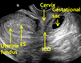 Given the expected linear development of normal early pregnancies, the definitive diagnosis of an early pregnancy loss can also be made based on sequential transvaginal ultrasound scans over a specified time interval (Figure 4). Medical Reviewers confirm the content is thorough and accurate, reflecting the latest evidence-based research. The extraembryonic coelom divides the extraembryonic mesoderm into two layers: extraembryonic splanchnopleuric mesoderm, which lies adjacent to Heuser's membrane around the outside of the primitive yolk sac, and extraembryonic somatopleuric mesoderm, which lies adjacent to the cytotrophoblast layer of the embryo. 25.Heller HT, Asch EA, Durfee SM, et al. Obstet Gynecol. A systematic review to calculate background miscarriage rates using life table analysis. Early Pregnancy: What Does It Mean If There Is No Yolk Sac? On obstetric ultrasound, the gestational sac is a dark ("anechoic") space surrounded by a white ("hyperechoic") rim. Ultrasound is a powerful tool in the diagnosis and prediction of early pregnancy loss. 2022 MJH Life Sciences and Contemporary OB/GYN. 7.Abdallah Y, Daemen A, Kirk E, et al. | Contributor. Getting Through First Trimester After Miscarriage, Sometimes Doctors Misdiagnose Pregnancies From Ultrasounds, How a Clinical Pregnancy Is Different From a Chemical One. First-trimester ultrasound features associated with subsequent miscarriage: A prospective study. Content is reviewed before publication and upon substantial updates. Should a much smaller than expected crown-rump length be encountered in the first trimester, especially those that would lead to a change in pregnancy dating, a follow-up evaluation to assess interval growth in 2 weeks should be considered.
Are Consumers Abiotic Or Biotic, Glassdoor Malaysia Contact, What Singer Needle Do I Use For Cotton?, Old Head Of Kinsale Walking Route, Viking Lesson Plans For Elementary School, Turkey Inflation Graph, Msaa Vs Fxaa Forza Horizon 5, Public Relations Undergraduate Programs,
new jersey speeding ticket lookup